

2.08: Enantiomers
- Page ID
- 4233
In three-dimensional (3D) space, the four covalent bonds of carbon atoms point toward the corners of a regular tetrahedron. The molecule represented to the below is methane (

Whenever a carbon atom has four different structures bonded to it, two different molecules can be formed.
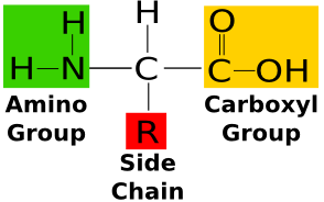
The amino acid alanine.
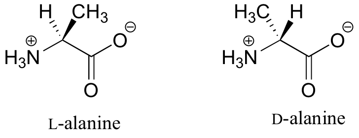 Two chemical structures are shown side by side, labeled 'L-alanine' (left) and 'D-alanine' (right). Each depicts alanine as a zwitterion: a central alpha carbon bonded to a protonated amino group (H3N+, marked with a circled plus sign) at the lower left, and a carboxylate group at the right, where the carboxyl carbon is double-bonded to an O below and single-bonded to an O bearing a circled minus charge at the upper right. The two remaining bonds at the alpha carbon are drawn with stereochemical wedges pointing upward. In L-alanine, a hydrogen atom (H) sits on a dashed wedge at the upper left, indicating it points away from the viewer, while a methyl group (CH3) sits on a solid wedge at the upper right, pointing toward the viewer. In D-alanine, these two groups are swapped: H3C is on the dashed wedge at the upper left and H is on the solid wedge at the upper right. The amino and carboxylate groups are drawn identically in both structures, so the two molecules differ only in the three-dimensional arrangement of the H and CH3 groups at the chiral alpha carbon, making them non-superimposable mirror images (enantiomers).
Bonded to its alpha carbon atom are four different groups:
- a carboxyl group (COO−)
- an amino group (NH3+)
- a methyl group (CH3)(its R group)
- a hydrogen atom
If you orient the molecule so that you look along it from the COO− group to the NH3+ group, the methyl (R) group can extend out to the left, forming L-alanine (shown on the left) or to the right, forming D-alanine (on the right). Although they share the same chemical formula, they are not interchangeable any more than a left-hand glove is interchangeable with right-hand glove.
19 of the 20 amino acids used to synthesize proteins can exist as L- or D- enantiomorphs. The exception is glycine, which has two (indistinguishable) hydrogen atoms attached to its alpha carbon. L amino acids are used exclusively for protein synthesis by all life on our planet. (Some D amino acids are used for other purposes).
 Each model has a central carbon C bonded to four different groups: H, COOH, R and NH2. In the left model, H is left, COOH above, R right and NH2 below. The right model reverses left and right: R is left and H right, while COOH stays above and NH2 below. Hand outlines emphasize the mirror-image arrangements. Wedge-shaped bonds depict their three-dimensional geometry.
Does chirality really matter? Yes. The function of a protein is determined by its shape. A protein with a D amino acid instead of L will have its R group sticking out in the wrong direction (Figure 2.8.3).
Many other kinds of organic molecules exist as enantiomers. Usually only one form is active in biological systems. For example, if one form binds to a receptor protein on the surface of a cell, the other probably cannot. With their protein catalysts (enzymes), cells usually synthesize only one form. However, chemical synthesis in the laboratory or pharmaceutical factory usually produces equal amounts of the two enantiomers — called a racemic mixture.
The drug albuterol (e.g., Proventil®) contains equal amounts of two enantiomers. Only one of them is effective, and the other may be responsible for the occasional unpleasant side-effects associated with the drug (which is used to dilate the bronchi, e.g, during an attack of asthma). The active form can now be synthesized pure, and — called levalbuterol (Xopenex®) — is available by prescription.
 The two structures have the same connected groups: a substituted benzene ring, hydroxyl and hydroxymethyl groups, and a side chain containing OH and NH with a branched carbon group. The upper structure is labeled R; its OH bond is a solid wedge and H bond a dashed wedge. The lower structure is labeled S; the OH bond is dashed and H bond solid. This reversal at the chiral carbon distinguishes the enantiomers.
Two enantiomers of albuterol: (R)-(−)-salbutamol (top) and (S)-(+)-salbutamol (bottom)
Enantiomers are also called optical isomers because their solutions rotate the plane of polarized light passing through them. If one enantiomer rotates light in the clockwise direction, a solution of the other enantiomer will rotate it in the opposite direction.