

4.07: Photosynthesis - Pathway of Carbon Fixation
- Page ID
- 4271
Photosynthesis is the synthesis of organic molecules using the energy of light. For the sugar glucose (one of the most abundant products of photosynthesis) the equation is:
Light provides the energy to transfer electrons from water to nicotinamide adenine dinucleotide phosphate (
The Steps that lead to Photosynthesis
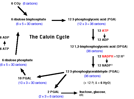 Flow diagram titled 'The Calvin Cycle' tracing carbon through photosynthetic carbon fixation; carbon counts appear in blue beneath each compound. At top left, 6 CO2 (6 carbons) feeds by arrow into 6 ribulose bisphosphate (6 x 5 = 30 carbons). An arrow leads right to 12 3-phosphoglyceric acid (PGA) (12 x 3 = 36 carbons). A downward arrow, with the side label '12 ATP' over '12 ADP', converts PGA to 12 1,3-bisphosphoglyceric acid (DPGA) (36 carbons). A further downward arrow, with the side label '12 NADPH + 12 H+' over '12 NADP+', reduces DPGA to 12 3-phosphoglyceraldehyde (PGAL) (36 carbons), annotated '(+ 12 Pi & + 6 H2O)'. From PGAL, one arrow points down-left to 10 PGAL (10 x 3 = 30 carbons); an upward arrow on the far left, labeled '6 ADP' over '6 ATP', carries 6 ribulose phosphate (6 x 5 = 30 carbons) back up to 6 ribulose bisphosphate, closing the cycle. A second arrow from PGAL leads down to 2 PGAL (2 x 3 = 6 carbons), which points right to the products 'fructose, glucose, etc.'. Net result: 10 of the 12 PGAL regenerate the CO2 acceptor ribulose bisphosphate, while 2 PGAL exit the cycle as carbohydrate output.
- CO2 combines with the phosphorylated 5-carbon sugar ribulose bisphosphate.
- This reaction is catalyzed by the enzyme ribulose bisphosphate carboxylase oxygenase (RUBISCO)(an enzyme which can fairly claim to be the most abundant protein on earth).
- The resulting 6-carbon compound breaks down into two molecules of 3-phosphoglyceric acid (PGA).
- The PGA molecules are further phosphorylated (by ATP) and are reduced (by NADPH) to form phosphoglyceraldehyde (PGAL).
- Phosphoglyceraldehyde serves as the starting material for the synthesis of glucose and fructose.
- Glucose and fructose make the disaccharide sucrose, which travels in solution to other parts of the plant (e.g., fruit, roots).
- Glucose is also the monomer used in the synthesis of the polysaccharides starch and cellulose.
The Figure 4.7.1 shows the steps in the fixation of carbon dioxide during photosynthesis. All of these reactions occur in the stroma of the chloroplast. These steps were worked out by Melvin Calvin and his colleagues at the University of California and, for this reason, are named the Calvin cycle.
Calvin's Experiment
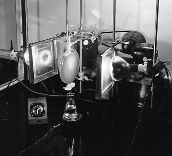 A flat, rounded glass vessel hangs vertically between lamps and square panels. Tubing, clamps and support rods surround it. A conical flask sits directly below, and a small dial instrument is at the lower left. The photograph is black and white; no instrument markings are readable.
The experimental apparatus is shown above. After various intervals of illumination, a suspension of unicellular algae is inactivated and the contents of the cells extracted. The compounds in a drop of the extract are then separated by paper chromatography.
The identity of each substance may be determined simply by comparing its position with the positions occupied by known substances under the same conditions. Or, a fragment containing the spot can be cut from the sheet and chemically analyzed.
To determine which, if any, of the substances separated on the chromatogram are radioactive, a sheet of X-ray film is placed next to the chromatogram. If dark spots appear on the film (because of radiation emitted by the 14C atoms), their position can be correlated with the positions of the chemicals in the chromatogram. Using this technique of autoradiography, Calvin found that 14C turned up in glucose molecules within 30 seconds after the start of photosynthesis. When he permitted photosynthesis to proceed for only 5 seconds, however, the radioactivity was concentrated in several other, smaller, molecules.
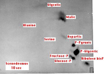 The image is labeled Scenedesmus 10 sec. Dark spots occur beside labels including Malic, P-Pyruvic, P-Glyceric and Ribulose bisP. Additional labels identify Glycolic, Alanine, Serine, Aspartic, Fructose-P and Glucose-P. P-Glyceric is a prominent lower-right spot. A small outlined rectangle appears at the bottom right. No numerical intensity scale is provided.
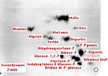 The label reads Scenedesmus 2 min. Dark spots are distributed among labels Malic, Alanine, Glycine, Glutamic, Citric, Serine, Aspartic, P-Pyruvic, Dihydroxyacetone-P, Ribose-P, P-Glyceric, Glucose-1,2-P, Ribulose bisP, Fructose-P, Sedoheptulose & Glucose-P, and Uridine di-P-glucose. Strong spots appear near Malic, Alanine and several phosphorylated compounds. A small outlined circle is at the lower right. No numerical intensity scale is shown.
The dark spots show the radioactive compounds produced after 10 secs (left) and 2 minutes (right) of photosynthesis by the green alga Scenedesmus. The alga was supplied with carbon dioxide labeled with 14C, a radioactive isotope of carbon. At 10 seconds, most of the radioactivity is found in 3-phosphoglyceric acid ("P-Glyceric"). At 2 minutes, phosphorylated 6-carbon sugars (glucose and fructose) have been synthesized as well as a number of amino acids. The small rectangle and circle (lower right-hand corners) mark the spots where the cell extract was applied.